ANGIOPOIETINS
IN RETINAL VASCULAR DISEASE
Scroll below to learn about the role of the angiopoietin/Tie2 pathway in retinal vascular diseases. First, learn how Ang-1 and Tie2 interact under normal conditions to achieve vascular stability and homeostasis. Next, learn how elevated Ang-2 can interfere with Tie2 signaling and lead to vascular instability, characterized by vascular leakage, neovascularization, and inflammation, contributing to
retinal vascular disease.
Together Ang-2 and VEGF-A play a critical role in the development of retinal diseases.
The interplay of Ang-2 and VEGF-A potentiates the progression of choroidal and
retinal vascular diseases.
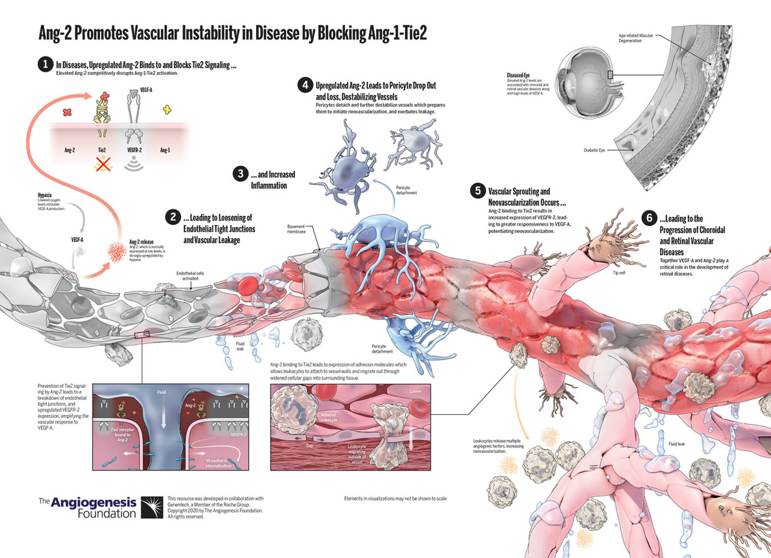
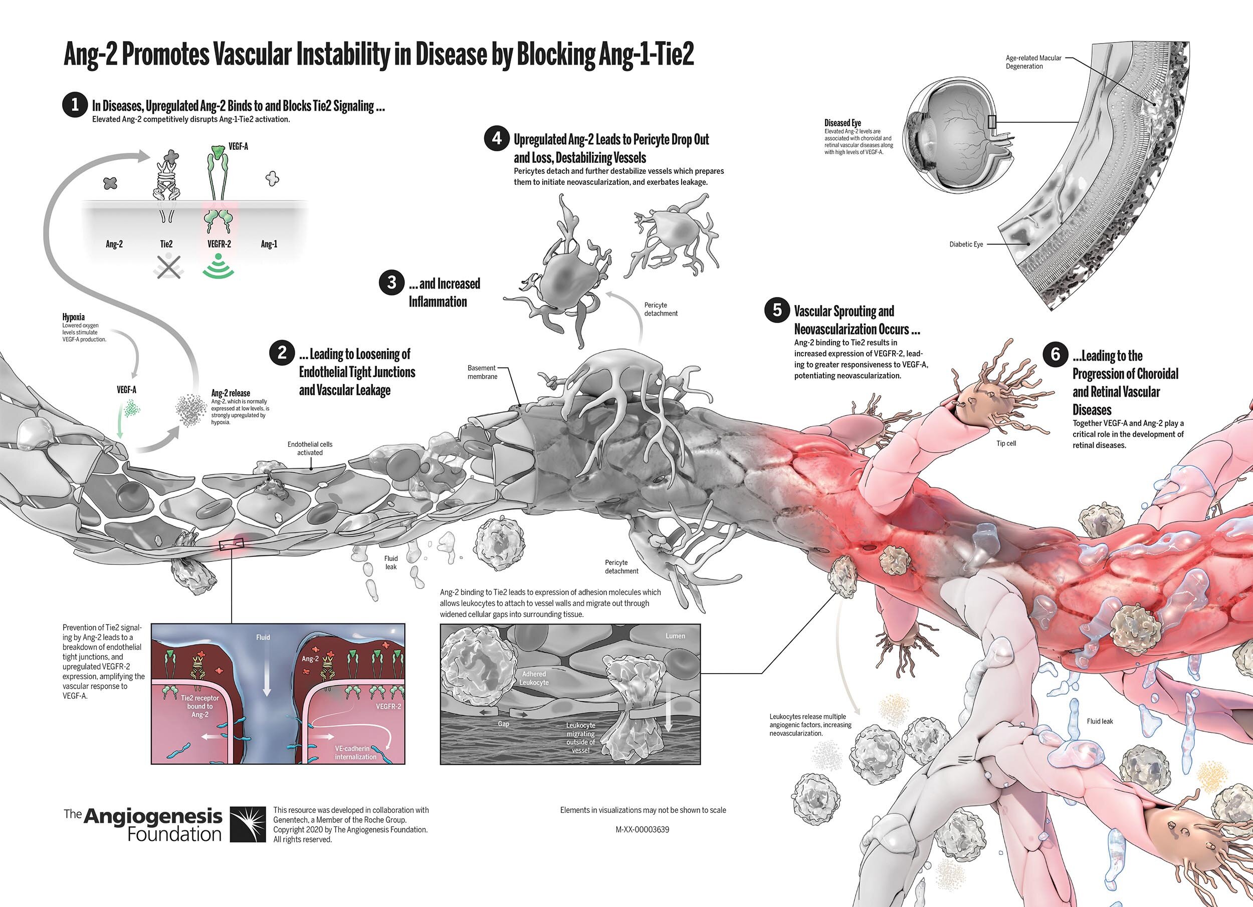
Below, the image shows the effect of Ang-2 + VEGF-A on a retinal blood vessel.
The greyed-out versions shows the effect of Ang-2 alone and
of VEGF-A alone, respectively.
Healthy retinal blood vessels are essential to eye health. Among their essential functions, they provide oxygen and nutrients to photoreceptors.
In healthy vessels, Ang-1 Promotes and Maintains Vascular Stability.
Ang-1 is made by platelets and pericytes and activates Tie2 receptors on endothelial cells to maintain vascular stability.
Ang-1 (Angiopoietin-1) is a vascular modulating growth factor that binds to an endothelial cell-specific tyrosine kinase receptor Tie2 and is responsible for vascular stability and maturation.
Tie2 is an endothelial cell surface receptor for
Ang-1 and Ang-2 responsible for intracellular signaling and intercellular attachment.
Ang-1-Tie2 signaling downregulates
VEGFR-2, thereby weakening VEGF responsiveness and preventing neovascularization.
Ang-1 activation of Tie2 leads to tightening endothelial cell junctions. Tightened cell-cell junctions prevent vascular leakage at the blood-retina barrier.
Downstream of Ang1-Tie2 signaling, pericytes are recruited for vascular stability and maturation.
As perivascular cells wrap around vessels, they inhibit endothelial proliferation and further reduce vascular leakage.
In healthy vessels, leukocytes flow through the bloodstream but do not adhere to vessel walls or transmigrate into the extracellular matrix.
Constitutive Ang-1-Tie2 signaling is critical for maintaining the non-proliferative, anti-inflammatory and non-angiogenic state of the healthy blood vessel. The result is endothelial quiescence
and homeostasis.
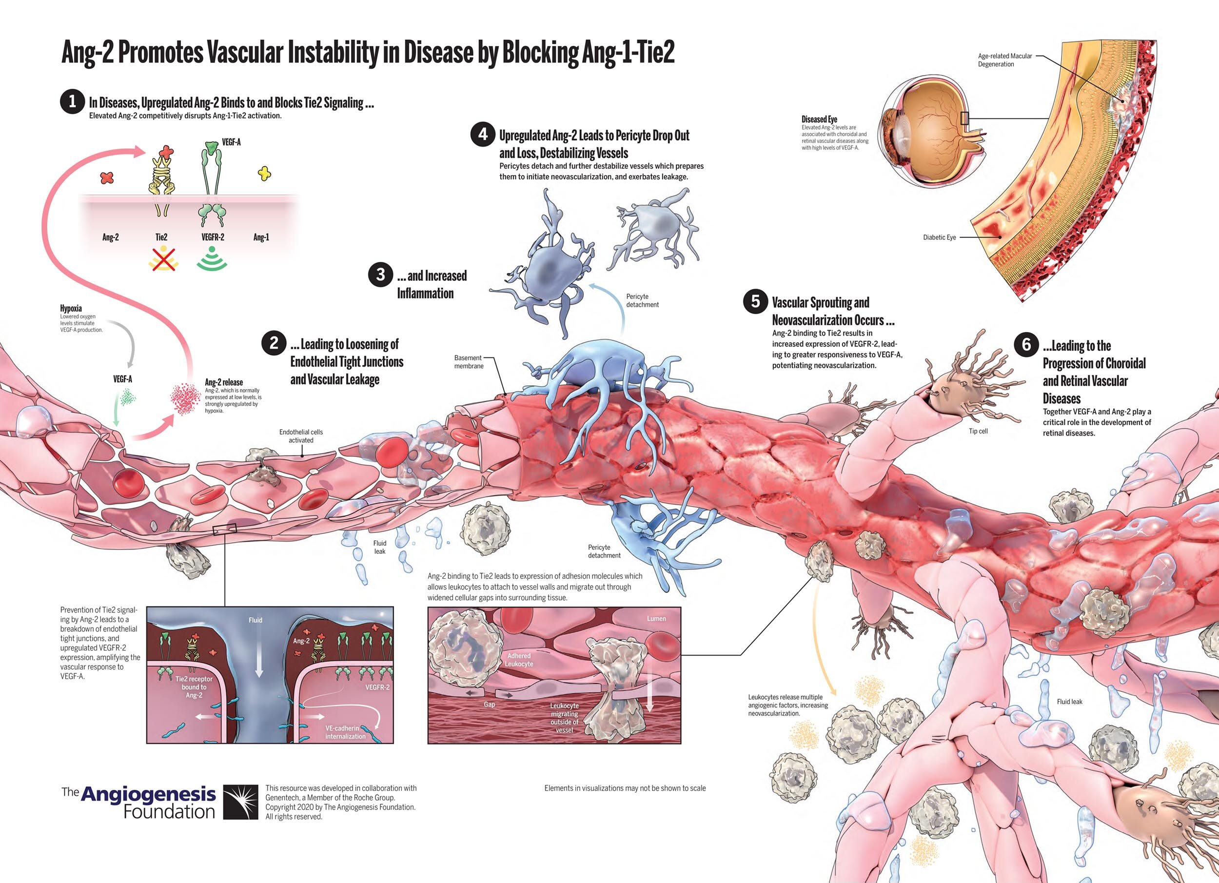
Ang-2 Promotes Vascular Instability in Disease by Blocking Ang-1-Tie2
In retinal diseases, upregulated Ang-2 binds to and blocks Tie2 signaling.
Prevention of Tie2 signaling by Ang-2 leads to loosening of endothelial tight junctions and vascular leakage, as well as upregulated VEGFR-2 expression, amplifying the vascular response to VEGF-A.
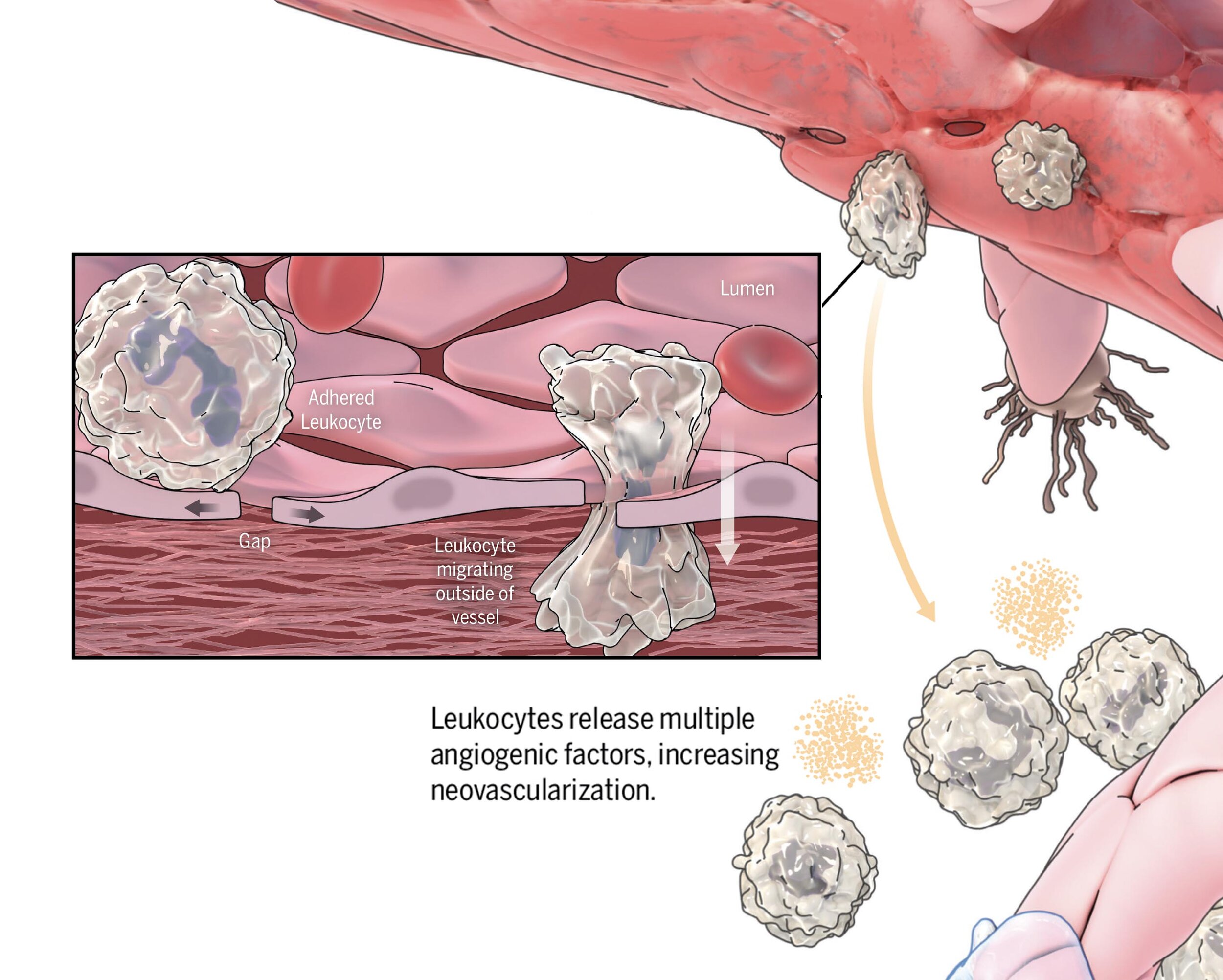
Inflammation occurs when Ang-2-Tie2 binding leads to expression of cellular adhesion molecules in endothelial cells. These molecules allow leukocytes to attach to vessel walls and migrate out through widened cellular gaps into surrounding tissue.
Upregulated Ang-2 leads to pericyte drop out and loss, destabilizing vessels. Pericyte detachment prepares vessels to initiate neovascularization, and exacerbates leakage.
Ang-2 binding to Tie2 results in increased expression of VEGFR-2 in endothelial cells.
This leads to greater responsiveness to VEGF-A, potentiating neovascularization.


